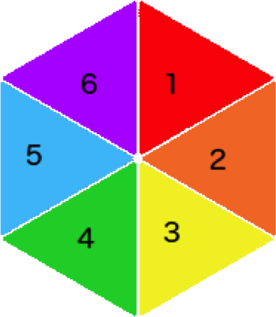
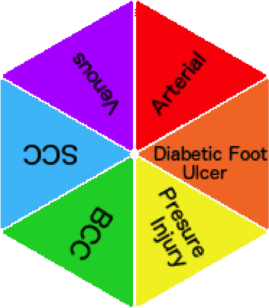
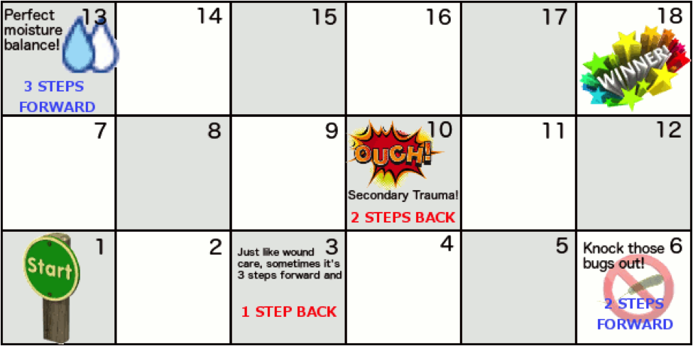
Cut out the two hexagons below and poke a toothpick through their centres to make two spinning tops. Spin the wound type top to determine your wound of choice, then spin the numbered top. You need to come up with that number of risk factors or signs that would lead you towards a diagnosis for that wound type. For example, if you had “Arterial” and “3” you could list; Edges-Punched out appearance, History-Smoker, History-Diabetes. Try to use all of HEIDI and TIME to help you come up with the answers and try not to duplicate! Place your piece on the starting square, if you’re successful, move your piece the same number of spaces. In my case it would be “3” spaces.


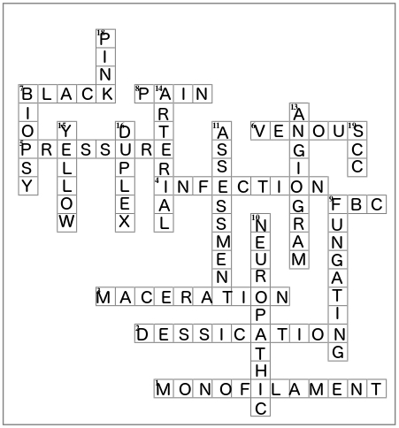
Across
- Tool most commonly used to assess sensation in feet.
- What happens to tissue when the wound is too dry
- What happens to tissue when the wound is too wet
- NERDS and STONES are two mnemonics to remember signs of...
- Wounds over boney prominences such as the sacrum and heels are often caused by ...
- These ulcers are most often found on the lower leg, are flat with irregular edges and highly exuding
- The colour most often associated with necrotic tissue
- Can be continuous, intermittent or procedural
- The blood test required to determine a white blood cell count
Down
- 10. These ulcers on the feet are caused by rubbing or trauma not felt by the patient
- 11. The most important part of the wound plan is following a systematic method of wound ...
- 9. Name given to cancerous wounds that have erupted beyond the normal borders of the skin
- 13. Investigation which displays an image of arteries or veins
- 14. The wounds tend to occur on extremities, over boney area, have a punched out appearance, painful ++
- 15. The colour often associated with slough
- 16. The best investigation for displaying venous reflux
- 7. Type of sample taken to best determine the presence of cancerous cells in a wound bed
- 18. The colour often associated with epithelial tissue
- 19. 90% of these non-melanoma skin cancers are found in sun-exposed areas of the body
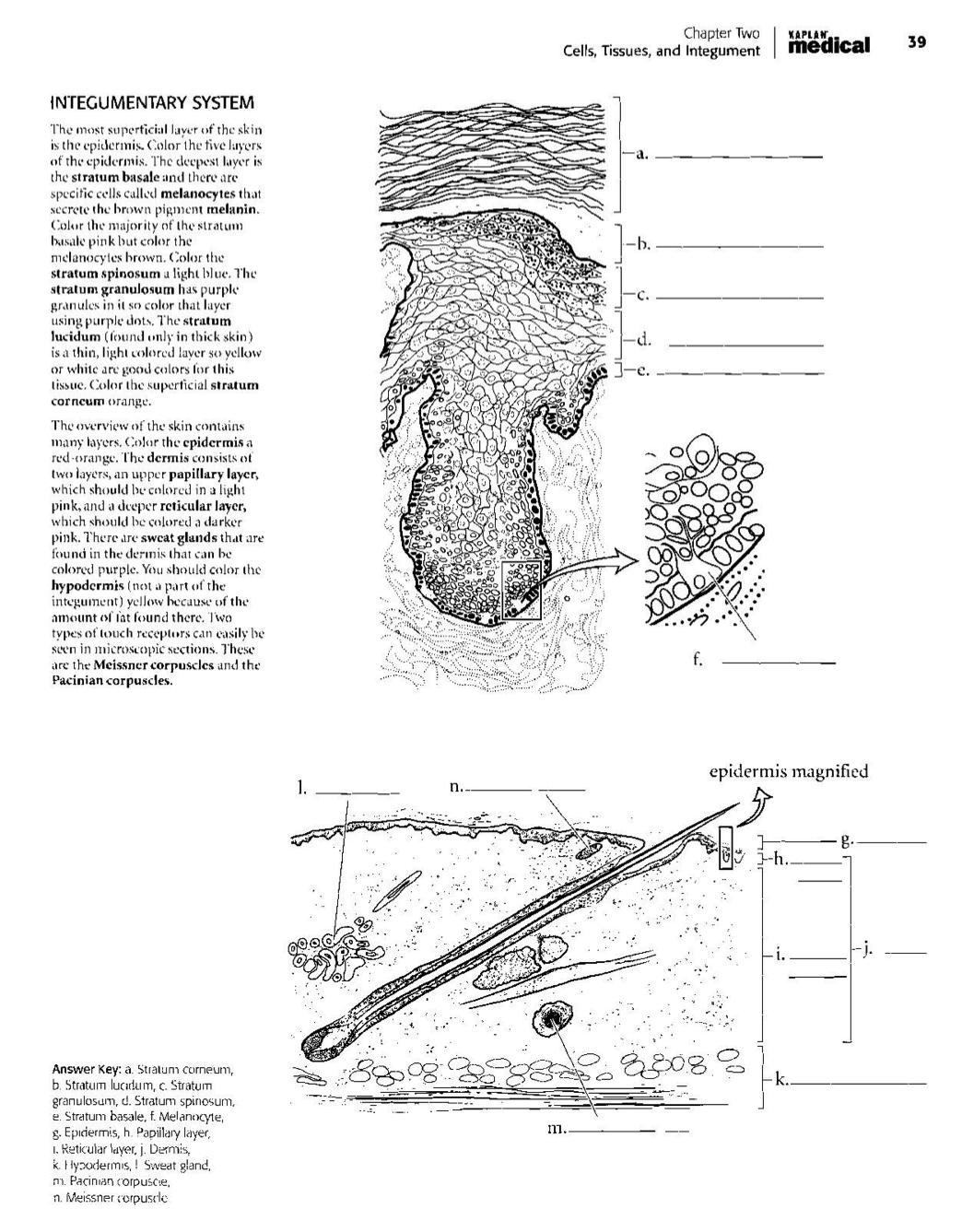
There are many available free at the Kaplan Anatomy Coloring Book